Seminar “Biomechanics as a driver of stem cell differentiation, segregation and development”
Date
Friday, 11th January 2018
Time
12:00 am
Place
University of Barcelona
Faculty of Physics Building
Room 3.20, 3rd floor
Speaker
Dr Lene Broeng Oddershede, Niels Bohr Institute (Germany)
Abstract
Early in embryonic development the blastocyst consists of a mix of two different cell populations, one primed towards the epiblast which will later develop into the fetus, and one primed for the endoderm, which will develop into the amniotic sac. Initially, those two populations are mixed and later they segregate into the fetus and amniotic sac. Using embryonic stem cells as a relevant model system, we use optical tweezers to quantify the physical properties of those two populations and demonstrate that their viscoelastic properties are significantly different. By modeling we also show that the difference in viscoelasticity in those two cell populations is actually enough to cause a segregation of the two populations, hence, the biomechanical properties alone can drive the process.
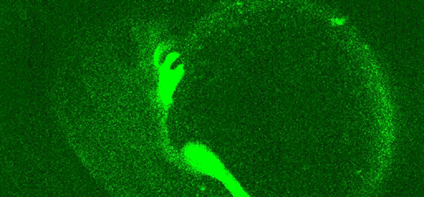
Mechanical forces and biophysical properties of cells are also vital for the morphogenesis of organs and embryos. However, how mechanical force and biophysical properties specifically contribute to tissue formation is poorly understood, predominantly due to a lack of tools to measure and quantify biomechanical parameters deep within living developing organisms without causing severe physiological damage.
Using an adapted version of optical tweezers we mechanically probe the developing gut region deep within living zebrafish and show that the mechanical properties of developing organs differ significantly. We hypothesize that these biomechanical differences serve as a segregation mechanism and might drive development.